XRAY CRYSTALLOGRAPHY STRUCTURAL BIOLOGY SHARED RESOURCE
at CU Cancer Center

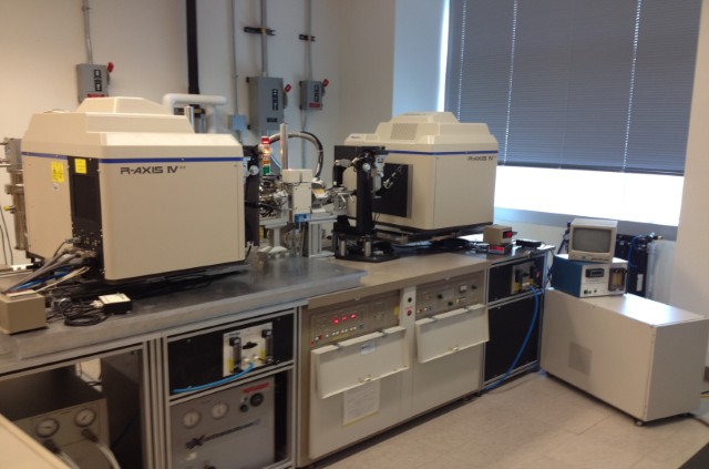
The X-ray crystallography facility is fully equipped for biomolecular crystallization, crystal screening, data collection, data processing, structure-determination and model building. The Rigaku/MSC Crystallization Robotics include: an Alchemist liquid handling robot for optimization screen production, a Phoenix drop setting robot, and 2 Desktop Minstrel imaging units, and a plate hotel that are all networked and integrated with Crystal trak software. The home source is composed of a Rigaku Micromax 007 high frequency microfocus X-ray generator, a VariMax high flux optic with adjustable divergence, an AFC11 4-axis goniometer, a Pilatus 200K 2D area detector, and an Oxford Cobra cryo-system. HKL-3000R is used for data collection and processing and is run on a Linux CentOS Dell Precision Workstation. The facility is located in RC1 South Building Rm 1301. It is directed by Dr. Mair Churchill (Department of Pharmacology), and is managed by John Hardin (Department of Biochemistry and Molecular Genetics).
Dr. Mair Churchill
Professor
Department of Pharmacology
CU Anschutz Medical Campus
12801 E. 17th Avenue, Mail Stop 8303
Aurora, CO 80045
Phone: 303-724-3670
Fax: 303-724-3663
Email: [email protected]
Dr. Jeffrey Kieft
Professor
Department of Biochemistry and Molecular Genetics
CU Anschutz Medical Campus
12801 E. 17th Avenue, Mail Stop 8101
Aurora, CO 80045
Phone: 303-724-3257
Fax: 303-724-3215
Email: [email protected]
Dr. Rui Zhao
Assistant Professor
Department of Biochemistry and Molecular Genetics
CU Anschutz Medical Campus
12801 E. 17th Avenue, Mail Stop 8101
Aurora, CO 80045
Phone: 303-724-3269
Fax: 303-724-3215
Email: [email protected]
Dr. John Hardin
Research Associate, Manager of the X-ray Core Facility
Department of Biochemistry and Molecular Genetics
12801 E. 17th Avenue, Mail Stop 8101
Aurora, CO 80045
Phone: 303-724-3330
Email: [email protected]
Professor
Department of Pharmacology
CU Anschutz Medical Campus
12801 E. 17th Avenue, Mail Stop 8303
Aurora, CO 80045
Phone: 303-724-3670
Fax: 303-724-3663
Email: [email protected]
Dr. Jeffrey Kieft
Professor
Department of Biochemistry and Molecular Genetics
CU Anschutz Medical Campus
12801 E. 17th Avenue, Mail Stop 8101
Aurora, CO 80045
Phone: 303-724-3257
Fax: 303-724-3215
Email: [email protected]
Dr. Rui Zhao
Assistant Professor
Department of Biochemistry and Molecular Genetics
CU Anschutz Medical Campus
12801 E. 17th Avenue, Mail Stop 8101
Aurora, CO 80045
Phone: 303-724-3269
Fax: 303-724-3215
Email: [email protected]
Dr. John Hardin
Research Associate, Manager of the X-ray Core Facility
Department of Biochemistry and Molecular Genetics
12801 E. 17th Avenue, Mail Stop 8101
Aurora, CO 80045
Phone: 303-724-3330
Email: [email protected]
007
Quick Start for 007- Turn on main N2 valve and let run for 5 minutes. Check the regulators: Cobra 1.0 mbar Pilatus 5 psi
- Turn on the power to the Pilatus and make sure that the power and temp lights on the back both turn green, can take up to 30 minutes. If the lights are not green check that the flow rate is at 15cc/min..
- If X-rays are off turn them on using the X-ray Generator Control software on the control computer. Under XG control turn X-rays on and then go to the Instrument menu and open XG script tool. Run MM007HFCuRampUp.xgr. Allow X-rays to come up to full power and run for at least an hour to get the best intensity possible. At the same time start up the cryo system (step 4).
- Turn on the power for the Cobra control box and run cool to –100K (about an hour).
- Turn on the crystal monitor.
- Close both doors to the generator room (Rm 1301) and turn on the Mag lock (on the pillar) and turn right/East safety interlock to “normal” position.
- Sign in the logbook noting filament hours, vacuum, and cold stream temperature.
- To protect the detector surfaces keep the cover on Pilatus while putting crystals up and down.
- When done mounting your crystal make sure the beam stop is clipped into place and Pilatus cover is removed.
- Start “HKL3000” and connect to the system under the collect tab. You are ready to do your experiments.
When done and if nobody is signed up to use the 007 within the next 48 hours:
- Turn of X-rays using the X-ray Generator Control software on the control computer. Under the Instrument menu open XG script tool. Run MM007HFCuRampDown.xgr. When script is done running turn X-rays off under XG control.
- Turn off the crystal monitor (label 5).
- Run End on Cobra control box (label 4 about 3.5hrs). When done turn off the Cobra Controller.
- Turn off the in house N2 using the main shutoff valve (label 1).
- Sign out in the logbook noting final fila
Safety Protocols
Radiation safety rules and the use of safety interlockBefore you go into the x-ray enclosure check that the safety interlock is on the “interlock” position.
General radiation safety rules:
- Always wear your body badge and ring badge when you need to go in the x-ray enclosure.
- Make sure all exposures are completed and the shutter is closed before you go in the x-ray enclosure.'
- Make sure the orange shutter light is off before you mount/dismount your crystals. It is always a good idea to keep an eye on this light when you work close to the direct beam.
- Never place any part of your body in the direct beam.
- Check the radiation with Geiger Counter whenever you are in doubt.
Available Software
- hkl (denzo and scalepack): Data processing. Current version 1.96.6.
- d*trek: Data processing. Current version DTREK72.
- Chooch: A program for deriving anomalous scattering factors from X-ray fluorescence spectra.
- CCP4: everything in crystallgoraphy except for data processing. Current version 4.1.
- Epmr: A molecular replacement program
- Replace: Another molecular replacement program
- Shelx: Structural determination and refinement for small molecule. Also useful in finding heavy atom positions as well as refinement of high resolution protein structures. Current version SHELX-97.
- Solve and Resolve: MIR/MAD structure determination and solvent flattening. Current version 2.01.
- CNS: structure determination and refinement. Current version 1.1.
- O: model building. Current version 8.0.
- Grasp: Graphical Representation and Analysis of Structural Properties
- Molscript and Bobscript: Graphical display of molecular 3D structures and electron densities.
- Ribbons: Graphical representation of molecular 3D structures.
The primary users of the X-ray facility include Drs. Mair Churchill (Pharmacology), Elan Eisenmesser (Biochemistry and Molecular Genetics), Jeffrey Kieft (Biochemistry and Molecular Genetics), David Jones (Pharmacology), Tanya Kutateladze (Pharmacology), and Rui Zhao (Biochemistry and Molecular Genetics). In addition, these primary users collaborate with Richard Davis (Biochemistry and Molecular Genetics), Michael Holers (Immunology), Bob Garcia (Pediatrics), and Heide Ford (Obstetrics & Gynecology) in a variety of research areas. Some recent publications by these users are listed below.
Churchill ME, Klass J, Zoetewey DL. Structural analysis of HMGD-DNA complexes reveals influence of intercalation on sequence selectivity and DNA bending. J Mol Biol 403(1):88-102, 2010 [PMC2962916]
Roy S, Musselman CA, Kachirskaia I, Hayashi R, Glass KC, Nix JC, Gozani O, Appella E, Kutateladze TG. Structural insight into p53 recognition by the 53BP1 tandem Tudor domain. J Mol Biol 398(4):489-496, 2010.
Hammond JA, Rambo RP, Filbin ME, Kieft JS. Comparison and functional implications of the 3D architectures of viral tRNA-like structures. RNA 15(2):294-307, 2009.
Liu W, Zhao R, McFarland C, Kieft J, Niedzwiecka A, Jankowska-Anyszka M, Stepinski J, Darzynkiewicz E, Jones DN, Davis RE. Structural insights into parasite eIF4E binding specificity for m7G and m2,2,7G mRNA caps. J Biol Chem 284(45):31336-31349, 2009.
Gangelhoff TA, Mungalachetty PS, Nix JC, Churchill ME. Structural analysis and DNA binding of the HMG domains of the human mitochondrial transcription factor A. Nucleic Acids Res 37(10):3153-3164, 2009 [PMC2691818]
Kieft JS. Comparing the three-dimensional structures of Dicistroviridae IGR IRES RNAs with other viral RNA structures. Virus Res 139(2):148-156, 2009 [PMC2673954]
Zhang L, Xu T, Maeder C, Bud LO, Shanks J, Nix J, Guthrie C, Pleiss JA, Zhao R. Structural evidence for consecutive Hel308-like modules in the spliceosomal ATPase Brr2. Nat Struct Mol Biol 16(7):731-739, 2009 [PMC2743687]
Champagne KS, Saksouk N, Pena PV, Johnson K, Ullah M, Yang XJ, Cote J, Kutateladze TG. The crystal structure of the ING5 PHD finger in complex with an H3K4me3 histone peptide. Proteins 72(4):1371-1376, 2008 [PMC2756976]
Laughlin, J.D., Ha T-S, Jones, D.N.M. and Smith, D.P. Activation of Pheromone-Sensitive Neurons is Mediated by Conformational Activation of a Pheromone-Binding Protein, Cell (2008) 133(7):1255-65.
Pfingsten JS, Constantino DA, Kieft JS. Structural basis for ribosome recruitment and manipulation by a viral IRES RNA. Science 314(5804):1450-1454, 2006
Chen Z, Zang J, Whetstine J, Hong X, Davrazou F, Kutateladze TG, Simpson M, Mao Q, Pan CH, Dai S, Hagman J, Hansen K, Shi Y, Zhang G. Structural insights into histone demethylation by JMJD2 family members. Cell 125(4):691-702, 2006
Shi X, Hong T, Walter KL, Ewalt M, Michishita E, Hung T, Carney D, Pena P, Lan F, Kaadige MR, Lacoste N, Cayrou C, Davrazou F, Saha A, Cairns BR, Ayer DE, Kutateladze TG, Shi Y, Cote J, Chua KF, Gozani O. ING2 PHD domain links histone H3 lysine 4 methylation to active gene repression. Nature 442(7098):96-99, 2006
English CM, Adkins MW, Carson JJ, Churchill ME, Tyler JK. Structural basis for the histone chaperone activity of asf1. Cell 127(3):495-508, 2006
Pena PV, Davrazou F, Shi X, Walter KL, Verkhusha VV, Gozani O, Zhao R, Kutateladze TG. Molecular mechanism of histone H3K4me3 recognition by plant homeodomain of ING2. Nature 442(7098):100-103, 2006
Charge Back Fee Schedule for X-ray Resource
| Service | Cancer Center Member-major user | Cancer Center Member | Non Cancer Member | Private Sector User |
| Service contract | $3000/yr | 0 | 0 | 0 |
| FacilityTraining Fee | $150 per person | $150 per person | $250 per person | $500 per person |
| Data Collection Usage | $20/hour (0-8 hours) 8-24 hr ($160) | $20/hour (0-8 hours) 8-24 hr ($200) | $30 per hour | $50 per hour |
| Crystallization | ||||
| Access per month | 0 | $100 | $150 | $200 |
| Alchemist | $12 | $15 | $25 | $35 |
| Phoenix | $12 | $15 | $25 | $35 |
| Materials/Labor | cost | cost | cost | cost |
| Crystal Screening | $300/half day | $300/half day | $500/half day | $1000/half day |
| Beamline usage | ||||
| Crystal screening | $30 ($3000 maximum) | $30 | $40 | N/A |
| Data set | $120 ($3000 maximum) | $120 | $160 | N/A |
*Price are subject to change
Access to facility by other faculty, staff, postdoc and students: Facility open to all CU Denver faculty, staff, postdocs and students, but need approval of the X-ray facility steering committee to arrange details.
Services Currently Provided
- Project design and consultation: Assistance with starting projects includes advice on protein expression and purification, and crystallization. Until now these services have been provided in direct collaboration with the primary users. Since the manager is now available, these services will be more broadly available to all members of the cancer center.
- Crystallization screening: Facilities for setting up crystal growth trays is available.
- Screening crystals: Crystal (diffraction) screening is available.
- Data collection: Data collection service is available.
Facility Managers
Dr. Mair Churchill
Professor, Director of the X-ray facilityDepartment of Pharmacology
University of Colorado Anschutz Medical Campus
12801 E. 17th Avenue, Mail Stop 8303
Aurora, CO 80045
Phone: 303-724-3670
Fax: 303-724-3663
[email protected]
Dr. John Hardin
Research Associate, Manager of the X-ray FacilityDepartment of Biochemistry and Molecular Genetics
12801 E. 17th Avenue, Mail Stop 8101
Aurora, CO 80045
Phone: 303-724-3330
[email protected]